Biology Journal Club
Giving an extra forum to Biology students.
In February 2026, one of our Biology teachers, Dr Bermudez, launched a new initiative: the Latymer Biology Journal Club. It is a weekly meeting where interested students will pick a research article, present it, discuss it, and dissect it. This will enable students to gain experience in preparing and delivering talks, whilst boosting their critical thinking skills.
Following their presentation, each student will write an article explaining their research, allowing them to develop their writing skills (no AI allowed – no exceptions). The resulting articles will be published below and will enable the readers to keep up-to-date with scientific advances.
Dr Bermudez - Why I created this club
I am sometimes asked by both staff and students alike why I switched careers from cancer research to teaching. My answer is straightforward and is always some variation of the following: because I love science, and because I find it very rewarding to work with young people to help them understand the world around us and develop a curiosity for it too (this was my favourite part of my PhD). By way of background, I do not come from a family of scientists or doctors, and when I moved from the US to London wanting to study science, I very much had to figure everything out on my own.
I am fortunate to have had some incredible mentors throughout my studies, like Professor Viji Draviam and Dr Louisa James at QMUL, and Professor Leonie Taams at KCL. They taught me some valuable life lessons that I try to carry forward and pass on to my own students. Among these is the importance of documenting the work you do. In a world where anyone can say that they’ve done anything, evidence is important, especially in a highly competitive environment like academia.
As an undergraduate student, I taught myself to code using the programming language ‘R’, and when I decided to host department-wide tutorials to help others learn too, Prof. Draviam said I should write an article about this and publish it via the QMUL newspaper. I followed her advice and was later able to reference this article when applying for PhD positions. I am certain that my following of this advice (for this situation and several others) was a key factor in my being awarded a Cancer Research UK funded PhD position, especially considering that I did not have a master’s degree and was competing against many who did!
This situation was brought to mind when a student I had previously taught for GCSE Biology, Shaan, came to my lab seeking advice on the anatomy of the hand. He was determined to 3D print a prosthetic hand at school, citing the massive costs for prosthetics. “What happens to people who cannot afford them?” he asked me. Shaan went on to spend the summer of 2025 working on this project, of his own accord (it was not done through a class at school, but instead in his free time), and when he was finished, he showed me the functional 3D printed hand and a PowerPoint he had created to explain the project. It worked – each finger could move independently, and the hand could grasp an object. Amazing. When I asked him what he was going to do with this work (Was he going to publish it somewhere? Surely, right?) I realised that he was going to reference it in his university applications, but that was it. Where was the evidence? I remembered Prof. Draviam’s words and relayed them to Shaan: “You need to publish this”. I sent some emails to scientific magazines; I did not hear back.
At the very same time, I was reading personal statements for my Year 13 students’ university applications. So many of them had done independent research and were applying for highly competitive places at universities with very little evidence to show for their independent efforts. And so was born my idea for the “Latymer Biology Journal Club”.
Let me explain. In universities and research institutes, a Journal Club is a well-established weekly meeting where one person presents a research paper and the group discusses it together. These meetings are an exercise in critical thinking combined with a desire to explore new research. I always really enjoyed them, partially because I would leave each one with a new perspective on the science, and partially because it made me realise that even research coming from the worlds’ leading scientists is not immune to constructive criticism. Things can always be improved. This should encourage us, not discourage us. Indeed, it is a very natural consequence of delving into the unknown world of scientific research. Something I notice regularly as a teacher, and especially in an environment like Latymer, full of bright and competitive students, is a certain reluctance to attempt something unless success is certain.
In the classroom, this looks like students not attempting to answer questions if they aren’t sure, or students being scared to offer suggestions, so on and so forth. But, to quote a brilliant article written by Martin Schwartz (The importance of stupidity in scientific research, Journal of Cell Science, 2008), “Focusing on the important questions puts us in the awkward position of being ignorant. No doubt, this can be difficult for students who are accustomed to getting the answers right. The more comfortable we become with being stupid, the deeper we will wade into the unknown and the more likely we are to make big discoveries.”
So here is my vision for the Latymer Biology Journal Club: a weekly meeting where interested students will pick a research article, present it, discuss it, and dissect it. Students will gain experience in delivering talks and improving their confidence when presenting. It will be a space to develop critical thinking skills and put them into practice, something that is especially crucial in today’s world, and something that I wish for them to carry with them long after they’ve left school. Journal Club will allow them to experience the discomfort of not always knowing the right answer in hopes of them realising that this not only okay, but actually essential in science. By having weekly meetings, they will be able to keep up to date with the latest scientific advances, which they may find useful during university applications.
Following their presentation, they will write an article explaining the research, allowing them to develop their writing skills (No AI – no exceptions). It will be published on the Latymer website, so that they can have evidence of this for university applications and beyond. This will also enable you, reader, to keep up to date with scientific advances. Finally, any student who develops their own research project, like Shaan, can discuss it with me and I will gladly give them a platform, for presenting and publishing their work. I believe in paying it forward, and that is what I am trying to do.
With 45 students currently signed up, it is my sincerest hope that you enjoy this series!
Shaan – Creating a 3D printed artificial hand
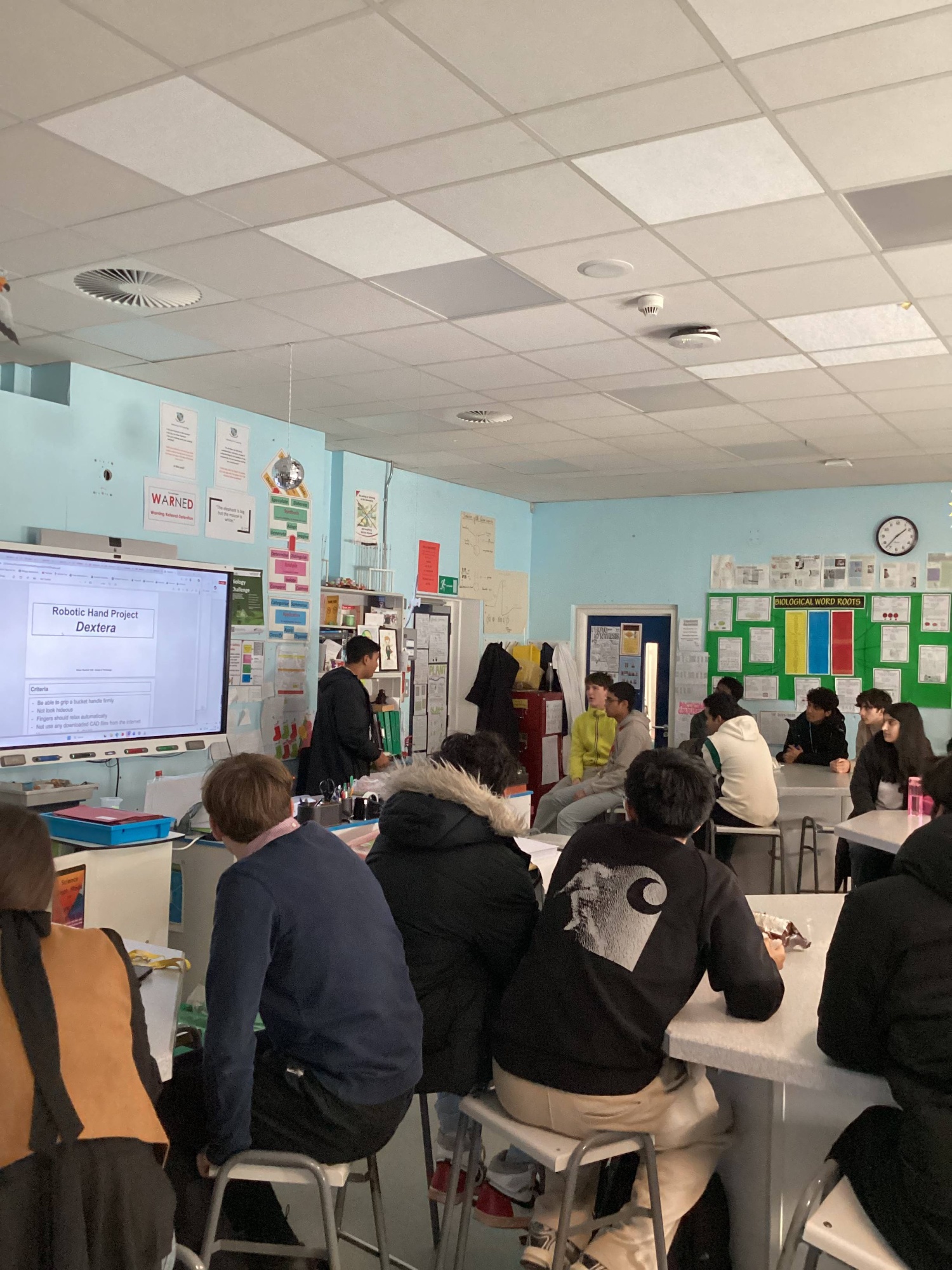 I had always looked at prosthetic arms and legs as an absolute necessity for people born without arms or legs, or having lost them in an accident. Some are lucky enough to have the opportunity to own an advanced prosthetic, but for those who do not have one, it is mostly due to their extortionate cost; one arm can cost upwards of £30,000. I didn’t believe that they should be that expensive, so I decided to see how expensive (and difficult) it would be to make one from scratch. "How hard can it be?”, I thought. As it turns out, very, very difficult.
I started out with a simple proof of concept of how tendons work to curl the fingers by 3D printing some trapezoidal prisms with holes in their centres and threading some old string through them. Having stuck them together with tape, I pulled the string and, lo and behold, it simulated the movement of the finger almost perfectly. This 10-minute experiment would go on to lay the foundation for this whole project; the concept of having a custom 3D printed finger being pulled by a string backwards is a deceptively simple task.
At this point, I began to experiment with the idea of a 3D printing concept called “Print in place”, where a dynamic mechanism can be printed in one piece and no assembly is required. This keeps the product as simple as possible and costs to a minimum. After 14 iterations of the finger, I had a decently successful working prototype. At this stage, in CAD (Computer Aided Design), I duplicated the fingers to create a 4-fingered hand. As wishy washy as it looked, it was able to function to an extent. I hooked it up to an Arduino Uno board and coded a loop programme for a servo motor to pull back the strings. It did not produce enough force to pull on all four strings at the same time, so it was back to the drawing board.
This time around, I chose to use four separate motors to operate the fingers. This brought its own challenges that I had to work my way around, such as power and control. Using 4x 180-degree servos with an auto-reset clause in the setup, I was able to make each move in the correct direction at the same speed with the same timings. However, this did not solve the issue of the fingers relaxing themselves, but for this I just stuck some elastic cord to the back as a temporary fix. Now I was able to control four fingers individually and reliably, the next task was to create an opposable thumb.
This was the biggest hurdle I had faced so far as there should be two axes of movement which were non-perpendicular. This meant I had to experiment a lot with different dimensions and angles in CAD, printing some examples throughout this process. Finally, I was able to add it to the design and print it out. I assembled it with all five motors and successfully ran a loop after some code fixes. The keyword here is loop. This whole product lacks the most important quality of a prosthetic; control.
I could have chosen a few different routes from here, but I decided to go with five momentary push buttons to operate the servos, on a push-to-actuate system. For this, I would have to get power, ground, and signal to all five buttons whilst maintaining a usable interface. Therefore, I designed a custom circuit board and soldered all the components and wires to a copper stripboard. With this board, I was able to design a housing for the electronics in CAD, and print it using PETG plastic. I bolted all the parts together and did some cable management, tested it, and at long last it worked! Each finger could be curled when the corresponding button was pressed.
Throughout the months I worked on this project, I was able to develop my CAD and 3D printing skills, having developed each component of the final product from scratch. More importantly though, I was able to achieve my goal from the beginning of determining whether prosthetics sold for £30k+ are fair practice.
The answer is no. My total development cost was £98, with a unit cost price of just £42. This is not to say that modern prosthetic hands aren’t more advanced, but rather to say that very similar technology can be developed for a tiny fraction of the cost, with the resources of a school. Every person who does not have a biological limb should have a basic robotic hand at the minimum, and this project goes to show it doesn't have to cost a literal arm and leg for such a necessity.
With special thanks to Mr. Collins, Head of Design & Technology.
Aswatha - Exploring the impact of T Cell research on immunotherapy
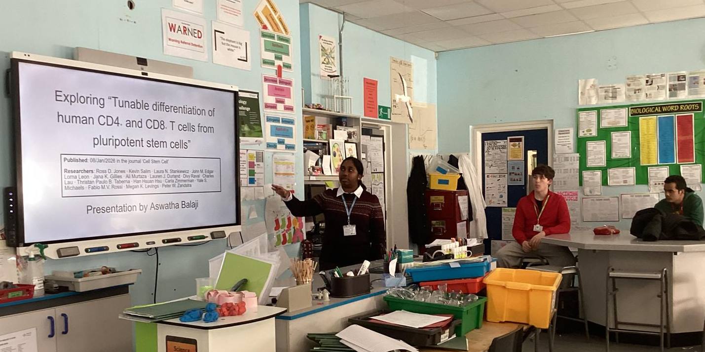 If you had asked me what I found most intriguing about the human body, I probably would have said our immune system. The idea that our bodies train specialised cells to patrol what are essentially the body's murky back alleys, spotting and eliminating suspicious figures within seconds, is something that still amazes me. These microscopic battles are happening constantly on so many fronts, yet we rarely notice any of it!
Seeing the role T cells played in both humoural and cell-mediated immunity, I began to question: what if, unlike CAR-T cell therapy, we could find a way to mature T cells outside of the patient's body? This would reduce the physical strain on the patient. What if, instead of relying entirely on adult donor-derived cells, we had a supply of prepared and nonspecific T cells at hand? lmmunotherapy could become quicker, cheaper, and thus, far more accessible!
In this study, researchers took pluripotent stem cells (PSC - these are embryonic cells with the natural potential to differentiate into almost any cell type) and attempted to differentiate them into human CD4+ and CD8+ T cells. While producing CD8+ T cells from PSC has already been demonstrated, it has been historically difficult to produce CD4+ T cells. At first glance, this may not seem like much of an issue since after all, it is the CD8+ T cells that directly destroy the tumour cells. However, CD4+ T cells sustain and coordinate the CD8+ T cells' immune response. This means that without them, the immune system's attack against the tumour is weakened. We can see this effect in diseases like HIV which specifically lower the patient's CD4+ T cell count, thus also making the patient vulnerable to opportunistic infections. If left untreated, the patient could develop AIDS later in life. Similarly, certain cancers and treatments, like
chemotherapy, can reduce the person's CD4+ T cell count. This makes being able to grow our own CD4+ T cells a groundbreaking advancement for treating cancer patients.
So, in order to generate CD4+ T cells, researchers of the "Tunable differentiation of human CD4+ and CD8+T cells from pluripotent stem cells" paper experimented with Notch signalling - a type of cell-to-cell communication where a ligand on the signal-sending cell binds to a Notch receptor on the signal-receiving cell. By carefully adjusting the signal's strength, they were able to control which corepressors were bound to the nucleus. This meant researchers could control which genes were expressed during the PSC's development, allowing them to successfully develop CD4+ T cells!
Even more impressively, these lab-grown cells were able to be polarised into the original four CD4+ T cell subtypes: Th1, Th2, Th17, Treg, demonstrating that we may no longer need to rely on adult donors to produce functional CD4+ T cells. Such an advancement would open the door to faster experimentation in T cell research. Perhaps different ratios of CD4+ and CD8+ T cells could be mixed to test which combination of the two would strengthen the weakened patient's immune response the most? The possibilities for further research using this development are endless and just the sheer number of them is incredibly inspiring.
On this happy note, I would like to thank Dr Bermudez and the school for giving me the opportunity to present an article on T cell research I found very fascinating!
Anjhana - Delving deeper into the mysteries of endometriosis
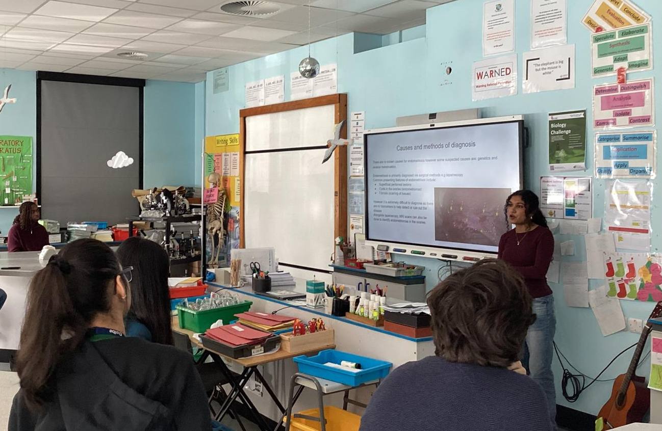 Imagine living with pain so severe it disrupts your daily life, yet doctors tell you consistently that it is just normal menstruation. Unfortunately, this is the reality for many women with endometriosis which is a chronic, inflammatory pain disorder where endometrial tissue is present outside of the uterus, most often in the pelvis. The reason why I decided to research endometriosis is because I was stunned by some of the statistics I found whilst reading up on it. It affects 1 in 10 women worldwide so around 190 million women are affected by it, however the average time for diagnosis is around 7-9 years. The question that I had was this: if it is such a common disease, then why does it take so long to get diagnosed?
Well, the most common symptoms of endometriosis are: chronic pelvic pain, dysmenorrhea (painful periods), dysuria (pain or discomfort when urinating), fatigue and infertility. There are no known causes of endometriosis however some suspected causes are genetics and reverse menstruation. Reverse menstruation is when blood flows upwards into the fallopian tubes instead of flowing out through the vagina. Several studies show that there is correlation between reverse menstruation and endometriosis as it is highly probable endometrial cells in the uterus make their way up into the pelvis through the fallopian tubes. The common presenting features are: superficial peritoneal lesions, endometriomas and fibrosis.
The primary method of diagnosing endometriosis is via laparoscopies as it is the only method of completely verifying that a person has endometriosis. MRI scans can also help diagnose it if they have cysts in their ovaries but it is not as effective of a method as laparoscopy. However, this is an issue as laparoscopy is an incredibly invasive method as it is surgical - the discovery of superficial peritoneal lesions via laparoscopy accounts for around 80% of diagnoses. This is one of the reasons why diagnosis is often delayed - not everyone can have laparoscopies and not everyone is willing to have them done. Additionally, the normalisation and minimisation of women’s pain and symptoms and a restricted access to care, especially in third world countries further exacerbate this issue.
This is why I was curious to see if there were any non-invasive methods of diagnoses being developed. Interestingly, a study has been carried out where the discovery of a saliva-based micro–ribonucleic acid (miRNA) signature for endometriosis in 2022 opened up new perspectives for early and non-invasive diagnosis of the disease. This is a non-invasive diagnostic tool that identifies small patterns of small non-coding RNA molecules in saliva to detect diseases. Some common diseases that this method is used for are: diabetes, HIV, oral cancer and cystic fibrosis. Specifically, the 109-miRNA saliva signature indicates that a person has endometriosis which acts as biomarkers which can be detected by using AI. They collected epidemiological data and saliva samples which were then analysed using Next-Generation Sequencing to profile genome wide miRNA expression by small RNA sequencing. They then fed this data into an AI model (random forest algorithm) which then determined whether or not the person had endometriosis.
Even if this is not a fully developed method yet, it still provides hope that there will be a non-invasive method of diagnosis in the future and the results of this study are truly inspiring to researchers. Overall, the issue of diagnosing such a common disease like endometriosis demonstrates a significant gap in the prioritisation of women’s health, emphasising the need for increased funding, research, and awareness to improve both diagnosis and treatment outcomes. Women should not be expected to tolerate chronic pain or endure years of delayed diagnosis for a condition that can have a profound impact on quality of life.
Finally, I’d like to thank Dr Bermudez for giving me the opportunity to present on a topic I am so passionate about!